Your doctor may order a variety of different diagnostic scans and tests to help monitor and diagnose your NHL. The variety of different scans is often confusing to patients, and many wonder why they are not getting one type of scan instead of another. The most important point to remember is that each type of scan has its purpose and place in treatment. No one scan type is superior to all the others because each serves a different purpose, and each is very valuable in its own right.
The first topic we will cover however is the recent (2014) recommendations about how and when to use PET/CT when diagnosing, staging and assessing the response for lymphoma.
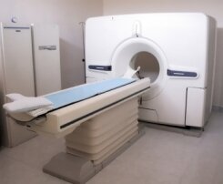
CT or Computed Tomography (often called CAT for Computed Axial Tomography) is the most common and useful type of scan for regular detection and follow up of NHL. CT's are still just X-ray images but they produce a near 3D image of the interior of the body. They do this by having the scanner spin around the body taking multiple pictures, or slices. A computer combines these images into a single image that looks like a slice taken right out of your body when looking down from the top of the head.
Click here to view a complete CT scan series
CT's are the most widely used scan because they allow the radiologist and doctor to look "inside" your body and see the structures that are there. They can see shape, size, and location of anything inside you. They provide far more detail than a simple x-ray image can. This is extremely important for finding tumours, and monitoring their growth, and seeing if they are threatening any nearby organs. Virtually all patients will undergo multiple CT scans throughout their journey with NHL. They are relatively inexpensive and give the doctors a great deal of information about what is going on. Even very small tumours can usually be spotted on a CT scan by an experienced radiologist. Since they have been around for a long time, there is a high degree of experience with them in the medical field so mistakes are less likely to happen.
Looking for more information about the various types of scans? There is a ton of it on the Internet. Just use your favourite search engine to search for "FDG PET", or "Gallium Scintigraphy" or "Computed Tomography". You'll find lots of information. One question that might occur to many patients is "Just how much radiation do I get from these various scans?" Of primary concern are X-rays, and CT's since they deliver radiation directly to your body. PET and Gallium scans inject radioactive substances into your body, but in only the most minute quantities. Nevertheless a PET scan delivers approximately the same amount of radiation to the body as a whole body CT scan.
It is a tough question to answer, but there is no doubt that a CT scan delivers dramatically more radiation to your body than any type of x-ray. A CT of the chest and abdomen can deliver the equivalent of 300 chest x-rays in radiation. While that sounds very frightening it is important to understand that this is still a small amount of radiation due to the very sophisticated equipment in use today. However if you are interested in reading a bit more about various radiation doses you get on a regular basis click the links below.
While not quite related to diagnostic imaging the following link looks at the risk of radiation from mobile phones which is still a concern to many people.
Mobile Phone Radiation and Health Guide
One question that is always on the mind of patients is, "how often do I need to have follow-up PET/CT scans one I have complete treatment?" There is no simple answer to that question. It depends a lot on the doctor and patient preference, as well as how well the patient responded to their treatment. If someone achieves a complete remission then follow-up scans might be less frequent than someone who was refractory to treatment. The need to know if the cancer is gone or has come back, has to be balanced with the risk of unnecessary radiation exposure.
To make this issue more confusing many recent studies have called into question the value of doing any surveillance scanning at all. Most of these studies have found that patients almost always experience symptoms of a relapse before any PET or CT scan finds it. This raises the question of whether or not we are doing scans that don't really need to be done, and exposing people to more radiation than necessary. Below are a variety of studies that look at this issue of surveillance scans in the follow up for lymphoma.